Imaging coupled vibrational, rotational, and electronic wave packet dynamics in a triatomic molecule
Abstract
Molecular dynamics triggered by interaction with light often involves the excitation of several electronic, vibrational, and rotational states. Characterizing the resulting coupled electronic and nuclear wave packet motion represents a severe challenge, even for small polyatomic systems. In this Letter, we demonstrate how the interplay between vibrational, rotational, and electronic degrees of freedom governs the evolution of molecular wave packets in the low-lying states of strong-field-ionized sulfur dioxide. Using time-resolved Coulomb explosion imaging (CEI) in combination with quantum mechanical wave packet simulations, we directly map the bending vibrations of the molecule, show how the vibrational wave packet is influenced by molecular alignment, and elucidate the role of the coupling between the two lowest electronic states of the cation. A conical intersection between these states couples the bending and asymmetric stretching coordinates, which is clearly reflected in the correlated fragment momenta. Our results suggest that multi-coincident CEI represents an efficient experimental tool for characterizing coupled electronic and nuclear motion in polyatomic molecules.
Recording molecular movies of an isolated molecule undergoing ultrafast structure changes with sufficient spatiotemporal resolution has been a longstanding goal in molecular optical sciences [1, 2, 3, 4]. Remarkable progress towards this goal has been demonstrated recently, including the development of gas-phase ultrafast electron diffraction (UED) [5, 6] and ultrafast X-ray scattering (UXS) techniques [7, 8, 9]. These techniques have provided novel insight into many photochemical reactions. However, the former still has limited time resolution, and the latter requires powerful X-ray sources due to low scattering cross sections. Both methods are insensitive to light atoms and often need access to large-scale facilities. They also project structural information (pairwise distances) onto a single coordinate (the momentum transfer), which makes unambiguous imaging of complex molecular dynamics difficult. Directly observing atomic motion during photochemical processes remains challenging [2, 4].
Another method used for making such movies is time-resolved Coulomb explosion imaging [10, 11, 12, 13, 14, 15, 16]. While CEI does not yield interatomic distances directly, it provides excellent temporal resolution, high sensitivity to light atoms, and accessibility in university-scale laboratories. Most importantly, coincidence imaging techniques [17, 18, 19, 20, 21, 22, 23] generate multidimensional CEI data that possesses an advantage in separating different reaction pathways, even for rare stochastic processes such as roaming [24].
Previously, all three methods have been employed to directly image light-induced vibrational wave packets (see, for instance, [19, 25, 26, 27, 8]), which play a critical role in many types of molecular dynamics [28, 29, 30, 31, 32, 33, 34, 35, 36]. However, most experimental movies of molecular vibrations have been limited to diatomic molecules (such as [37, 19], [38, 39], [40], [41, 40], [42, 40], and [25, 26, 27]) or the stretching of only one bond in polyatomic molecules [43, 44, 45]. Due to the fast timescale and complexity of rovibronic motions in polyatomic molecules, only a limited number of direct structural measurements of other vibrations has been reported [46, 47, 8].
In this Letter, we demonstrate that laser-induced Coulomb explosion can directly image ultrafast coherent wave packets in a small polyatomic molecule and produce movies that show the motion of individual atoms even in the presence of coupled nuclear and electronic motion. We investigate sulfur dioxide (SO2), a bent triatomic molecule that has been studied extensively (see, for instance, [48, 49, 18, 12, 50, 51, 52, 53, 54, 55, 56]) due to its significance in atmospheric processes and its intriguing photochemistry, including the efficient formation of neutral and ionic molecular oxygen [57, 58, 59, 60]. We first demonstrate the use of pump-probe CEI and coincident measurement of ion momenta to image the coherent vibrational wave packet in SO generated by ionization. These observations are supported by quantum mechanical wave packet calculations and classical Coulomb explosion simulations. We then show that, although recoil-frame ion momenta best represent this vibrational wave packet, laboratory-frame momenta reveal the importance of rotational motion in determining the overall dynamics. Finally, we show that a secondary wave packet resulting from a resonant dipole coupling between two ionic states traverses a conical intersection and leaves signatures of the nonadiabatic coupling in the ion momentum distributions.
Our experiment utilized a Ti:Sapphire laser operating at 10 kHz, generating near-infrared (NIR) pulses with a central wavelength of approximately 790 nm and a pulse duration of about 28 fs. We split this output into pump and probe pulses, with independently controlled power, and scan their relative delay using a motorized linear stage. As depicted in Fig. 1, we use a 3.4 W/cm2 pump pulse to ionize SO2 and a 8 W/cm2 probe pulse to further ionize and dissociate (i.e., Coulomb explode) it. The ions are then detected in coincidence using a COLTRIMS apparatus [61]. The momenta of these ions, determined from their measured time of flight and impact position, are used to deduce information about the induced wave packets.
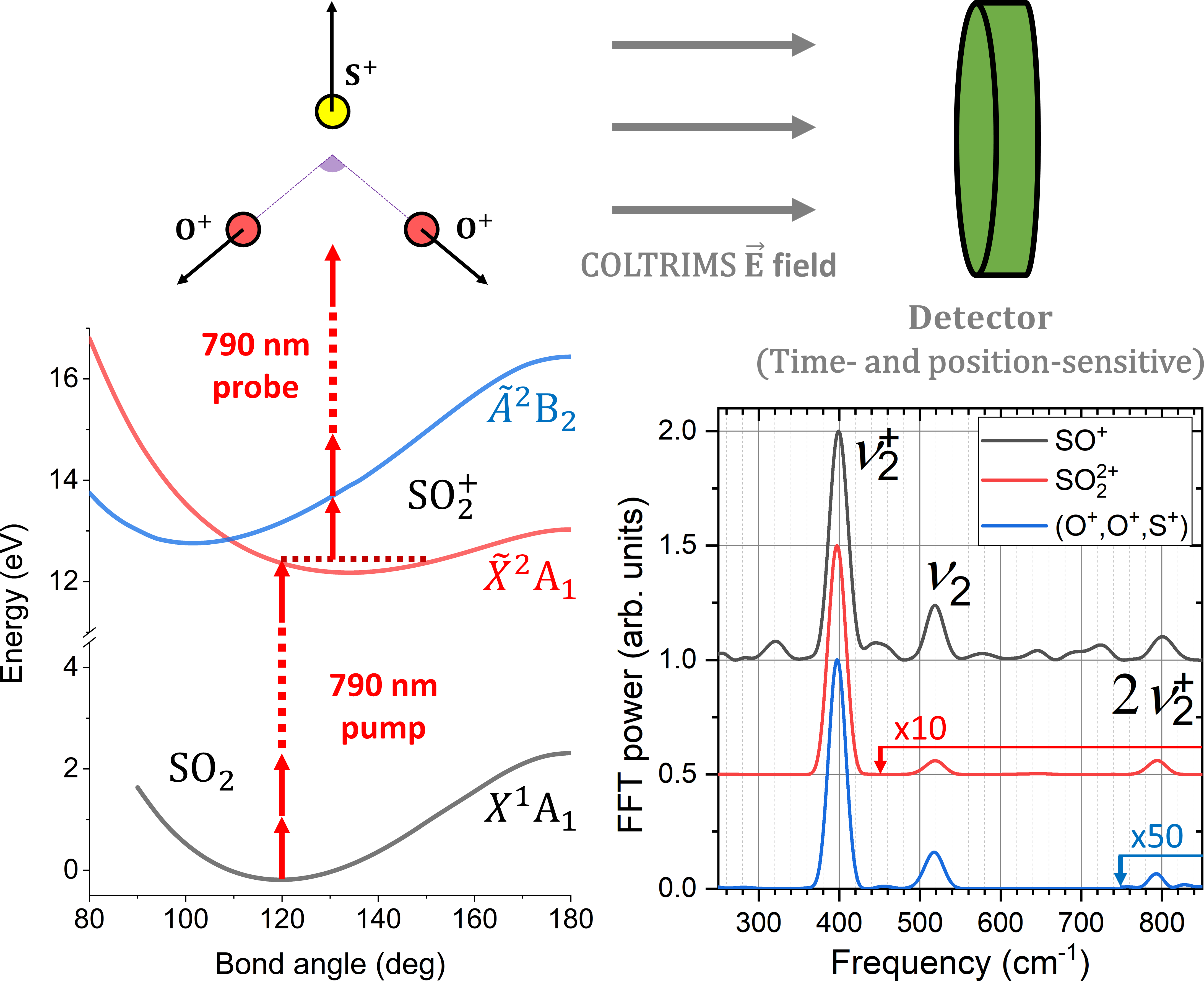
Previous studies [62, 63, 64, 65] have reported that the bending mode progression dominates the SO () SO2 () photoelectron spectra because the equilibrium geometries in the electronic ground state of the neutral SO2 () and ionic SO () have almost identical bond lengths but different bond angles [62, 66]. This leads to a strong excitation into the bending mode () and very weak excitation into the symmetric ( and asymmetric () stretch modes. The first seven bending-mode energies are approximately equally spaced with a nearest-neighbor separation of cm-1. In our experiment, which has a 17 cm-1 frequency resolution, oscillations at this frequency (and its first overtone at cm-1) are observed in different observables for various ionization and fragmentation channels. This indicates that these channels involve the population of SO ground state by the pump pulse and subsequent ionization and/or fragmentation by the probe. For example, as shown in Fig. 1, the fast Fourier transform (FFT) spectra of the delay-dependent yields of SO+ and SO ions and the three-body triply-charged final-state channel show a strong peak at the bending frequency and a much smaller one at the overtone. They also show a weak but clear signature of the bending vibration in the ground state of neutral SO2 (at 520 cm-1) [67]. In this Letter, we focus on the three-body channel to directly visualize the bending vibrations of the cation via delay-dependent kinetic energy (KE) spectra and angle correlations between the fragment ions.
In Fig. 2, we present experimental data from the channel illustrating the delay dependence of several key observables: (a) the angle between the momentum vectors of two O+ ions [ ], (b) the kinetic energy of S+ fragment [KE()], and (c) the total kinetic energy of two O+ fragments [KEsum()]. Simulation results for these observables are depicted in panels (d-f) and their delay-dependent mean values are shown in panel (g). Panel (h) features distributions of the ions at 250 fs (scatter plot) and 210 fs (contour line) when the bending wave packet is at the inner and outer turning points, respectively.
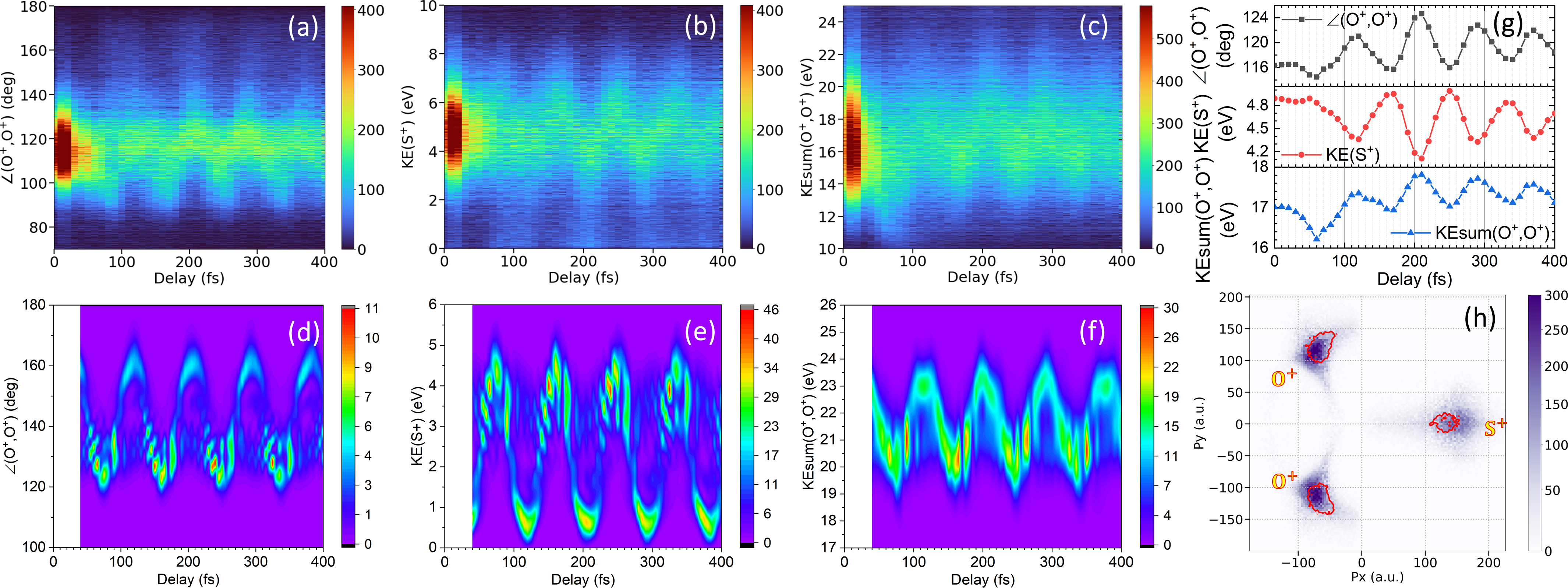
There are two distinguished features in the experimental data shown in Fig. 2(a-c). First, all the observables, namely , KE(), and KEsum(), show pronounced oscillations. The periodicity of these observed oscillations is about 83 fs (or 400 cm-1), clearly indicating that we mainly image the vibrational wave packet in the ground state of SO [63]. Second, there is a clear correlation between these observables: and KEsum() oscillate in-phase with each other and out-of-phase with KE(S+). This can be explained in terms of Coulomb forces between point charges (see Fig. S3 in the SM): near the equilibrium geometry, the net force on the S+ (O+) fragment due to the other two decreases (increases) with increasing bond angle. Moreover, also increases monotonically with the real-space bond angle.
We numerically model the experimental observables in Figs. 2(a-c) by solving the time-dependent Schrödinger equation for the coupled nuclear motion on the Born-Oppenheimer potential energy surfaces of the SO2 () and SO () electronic ground states, including all vibronic degrees of freedom (symmetric stretch, bending, and antisymmetric stretch modes) [74]. We calculated the molecular electronic states ab initio by applying the multi-configurational self-consistent-field method as implemented in the quantum chemistry code GAMESS [75], based on seven frozen inner orbitals and 12 active orbitals expanded in the correlation consistent-polarized valence triple zeta (cc-pVTZ) basis set. Following the dipole coupling of the nuclear motion in the neutral and ionic ground states by the pump pulse, we model the delayed probe pulse by vertically projecting the nuclear motion onto the potential energy surface of the triply-charged molecule, approximated as purely Coulombic, and propagate the nuclear wave packet to sufficiently large internuclear distances on the (O+, O+, S+) asymptote to achieve numerical convergence. We obtain the fragment kinetic energy releases by Fourier transformation of the real-space nuclear wave function to momentum space [76].
Our simulations in Figs. 2(d-f) closely resemble the experimental data in Figs. 2(a-c), manifesting similar periodicity, phases, and correlations between the delay-dependent fragment angles and fragment kinetic energies KE() and KEsum(). However, we note that each pair of experiment-simulation plots shows a mismatch in absolute value because vertical projection to and propagation on the Coulomb potential do not adequately describe the ionization and fragmentation process caused by the probe pulse. Nevertheless, these results provide a direct intuitive picture of the strong-field-induced vibrational wave packet motion in a triatomic molecule. Modulations of this three-body channel are dominated by the ionic ground-state vibrations, with a weak contribution from bending vibrations in the ground state of neutral SO2 as revealed by Fourier transformation of the delay-dependent data (see Fig. 1 and Fig. S2 of the SM).
Besides revealing the states involved in the vibrational wave packet, coincidence momentum imaging also allows us to visualize the correlated motion of individual atoms. To achieve this, we plot the data for all ions from the channel in a 2D momentum image (Newton plot). Fig. 2(h) features a representative image at 250 fs with a contour for data at 210 fs (the inner and outer turning points of the bending wave packet). Because the and KE() manifest the same behavior as the OSO bond angle and the displacement of S from the center of mass (see Fig. 2 and Fig. S3), movies comprising a timed sequence of these images (as provided in the SM) are a striking and intuitive representation of the bending vibration of the molecule. These molecular movies illustrate that during the first 400 fs, the wave packet is well localized and mimics the motion of a “ball-and-stick” model.
While bending vibrations are responsible for the most pronounced features of the observables shown in Figs. 1 and 2, the experimental data contain more information about the molecular wave packet launched by the pump pulse. One hint of other dynamics is found in the total yield of the (O+, O+, S+) channel, which does not exhibit oscillations at the frequency of the bending vibration. This suggests that double ionization probability of the cation is insensitive to this mode. However, as depicted in Fig. 3(a), the yield initially decreases by approximately 15% within the first few hundred femtoseconds before flattening out. To explain this behavior, we also plot for this channel in Fig. 3(a). Here, is the angle between the laser polarization and , representing the direction of the O-O axis. The strong correlation between these two quantities (except near the overlap region) indicates that the observed yield variation is due to the initial alignment of the pump-induced rotational wave packet. The time scale is consistent with the rotational dynamics of the two-body breakup channels shown in Fig. S4 (SM). Although the O-O axis is the most polarizable axis of the molecule, strong-field ionization of neutral SO2 is more likely to occur along the symmetry axis (see [54] and SM, Fig. S5). Thus, SO ions are most likely created near the peak of the pump pulse with their symmetry axes aligned with the laser polarization. They then rotate due to the angular momentum accumulated in the pump pulse and reach peak alignment (of the O-O axis) around 200 fs. After the peak, the rotational wave packet dephases and exhibits long-term incoherent alignment.
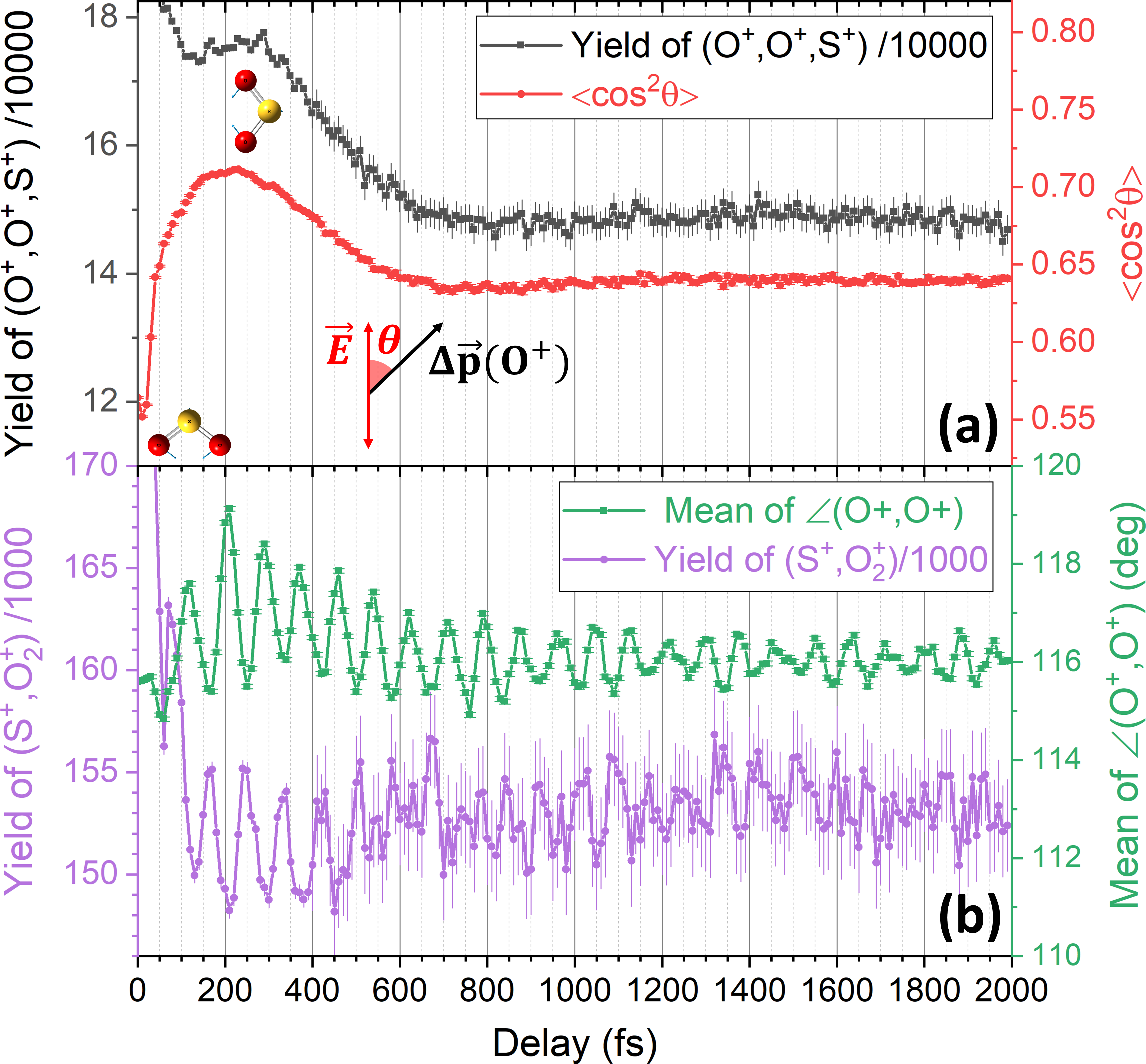
This rovibrational wave packet motion in SO has other manifestations that can be elucidated by multi-coincidence momentum imaging. One example is the formation of O, which can be traced by analyzing the (S+,O) coincidence channel. The delay-dependent yield of this channel is shown in Fig. 3(b) along with the mean value of in the three-body channel. Clearly, the two oscillate out of phase, suggesting an intuitive picture of O formation by the probe pulse: O is more likely to be formed if the probe pulse finds the cation at smaller OSO angles, where the two oxygen atoms are closer to each other. The alignment and bending angle dependence of O production may help improve our understanding of a potential source of abiotic oxygen in SO2-rich planetary atmospheres [58].
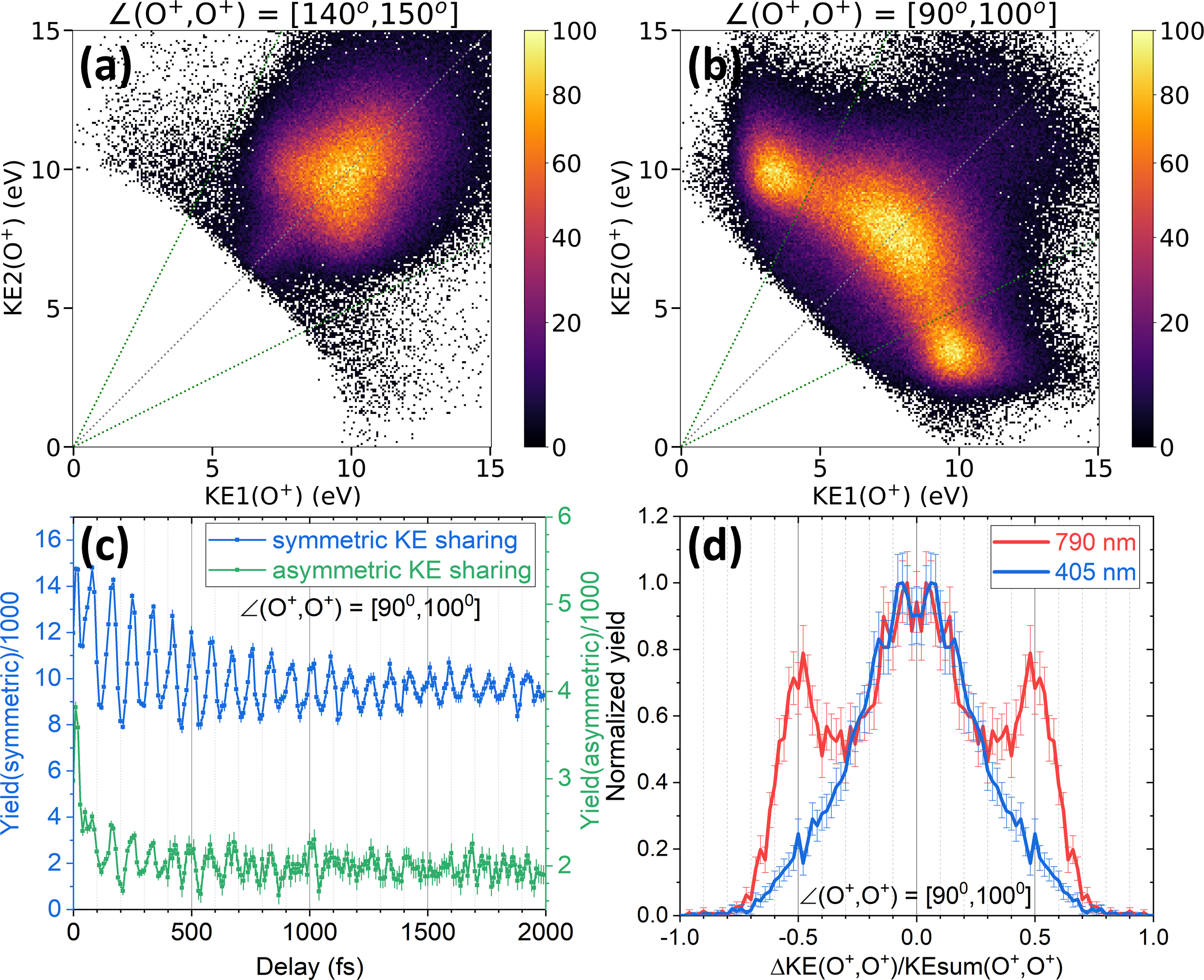
The rovibrational wave packet also controls the excitation of the ground-state cations to the first excited state in the probe pulse. This single-photon transition is energetically allowed for and its dipole moment is along the O-O axis. Thus, when ground state cations have rotated into alignment with the probe laser polarization and bending mode vibration has pushed the nuclear probability density to smaller-then-equilibrium bond angles (i.e., ), a single photon in the leading edge of the probe can resonantly transfer population from to (see Fig. 1 for PE curves). These two states have a symmetry-allowed conical intersection (CI) at with the branching space comprised of the bending and asymmetric stretching coordinates [53]. After excitation to the wave packet travels towards the CI where it is nonadiabatically coupled to the asymmetric stretch mode and returns to within 20 fs [53]. Since our probe pulse width is 28 fs, these cations can still be ionized at the peak of the probe and appear in the (O+, O+, S+) channel. On the other hand, if the excitation occurs in the trailing edge of the pump pulse, the wave packet should dephase rapidly as it traverses the CI and not exhibit periodic oscillations at 400 cm-1.
In the experimental data, signatures of a wave packet in state are seen in the KE sharing plot for the O+ ions. We separate events with and in Fig. 4(a,b), respectively, with the expectation that the population excited to can travel to smaller bending angles than the inner turning point of bending wave packet in but not to the outer turning point within the probe pulse. The strong component with asymmetric KE sharing seen in Fig. 4(b) but not in Fig. 4(a) confirms the dipole coupling to and nonadiabatic coupling to asymmetric stretch mode. Fig. 4(c) shows the delay-dependent yields of events with symmetric (blue) and asymmetric (green) KE sharing [all with small ]. The in-phase oscillation of the two curves is consistent with population transfer by the probe to the SO () state near the inner turning point of the ground state bending vibration. To further test this hypothesis of resonant inter-state coupling, we use a single laser pulse at 405 nm to ionize SO2 into (O+, O+, S+). The absence of asymmetric KE sharing peaks in the 405-nm case [Fig. 4(d)] confirms a resonant coupling near 790 nm, corroborating our conclusion that we see signatures of mode-coupling at the CI in our pump-probe experiment.
In conclusion, our work highlights the direct sensitivity of CEI to changes in the spatial density of the nuclear wave packet, demonstrating the feasibility of tracking the correlated motion of individual atoms within a molecule. This approach offers intuitive mechanistic insights into various molecular dynamics. The high-dimensional measurement allows us to observe and interpret the interplay between rotational, vibrational, and electronic motion in determining the molecular wave packet evolution and the experimental outcomes. Our simulations successfully capture the periodicity and phase behavior of different observables and their correlations, strongly supporting our interpretations. In view of recent advancements demonstrating that CEI can image detailed 3D structures of gas-phase molecules with about ten atoms [21, 23], this work paves the way for tracking dynamics of medium-size polyatomic molecules with unprecedented detail.
Acknowledgements.
We thank Charles Fehrenbach for taking care of PULSAR laser operation. We are grateful to the technical staff of the J.R. Macdonald Laboratory for their support. This work and the operation of the J.R. Macdonald Laboratory are supported by the Chemical Sciences, Geosciences, and Biosciences Division, Office of Basic Energy Sciences, Office of Science, U.S. Department of Energy, Grant no. DE-FG02-86ER13491. The PULSAR laser was provided by Grant no. DE-FG02-09ER16115, S.B. is supported by Grant no. DE-SC0020276 from the same funding agency, A.S.V. by the National Science Foundation Grant no. PHYS-1753324. S.J. was funded by the RISE Worldwide program from the German Academic Exchange Service (DAAD) for her summer research at Kansas State University.References
- Zewail [2000] A. H. Zewail, Femtochemistry: Atomic-Scale Dynamics of the Chemical Bond, The Journal of Physical Chemistry A 104, 5660 (2000).
- Ischenko et al. [2017] A. A. Ischenko, P. M. Weber, and R. J. D. Miller, Capturing Chemistry in Action with Electrons: Realization of Atomically Resolved Reaction Dynamics, Chemical Reviews 117, 11066 (2017).
- Ivanov [2021] M. Ivanov, Concluding remarks: The age of molecular movies, Faraday Discuss. 228, 622 (2021).
- Zhang et al. [2022] M. Zhang, Z. Guo, X. Mi, Z. Li, and Y. Liu, Ultrafast Imaging of Molecular Dynamics Using Ultrafast Low-Frequency Lasers, X-ray Free Electron Lasers, and Electron Pulses, J. Phys. Chem. Lett. 13, 1668 (2022).
- Weathersby et al. [2015] S. P. Weathersby, G. Brown, M. Centurion, T. F. Chase, R. Coffee, J. Corbett, J. P. Eichner, J. C. Frisch, A. R. Fry, M. Gühr, N. Hartmann, C. Hast, R. Hettel, R. K. Jobe, E. N. Jongewaard, J. R. Lewandowski, R. K. Li, A. M. Lindenberg, I. Makasyuk, J. E. May, D. McCormick, M. N. Nguyen, A. H. Reid, X. Shen, K. Sokolowski-Tinten, T. Vecchione, S. L. Vetter, J. Wu, J. Yang, H. A. Dürr, and X. J. Wang, Mega-electron-volt ultrafast electron diffraction at SLAC National Accelerator Laboratory, Review of Scientific Instruments 86, 073702 (2015).
- Filippetto et al. [2022] D. Filippetto, P. Musumeci, R. K. Li, B. J. Siwick, M. R. Otto, M. Centurion, and J. P. F. Nunes, Ultrafast electron diffraction: Visualizing dynamic states of matter, Rev. Mod. Phys. 94, 045004 (2022).
- Minitti et al. [2015] M. P. Minitti, J. M. Budarz, A. Kirrander, J. S. Robinson, D. Ratner, T. J. Lane, D. Zhu, J. M. Glownia, M. Kozina, H. T. Lemke, M. Sikorski, Y. Feng, S. Nelson, K. Saita, B. Stankus, T. Northey, J. B. Hastings, and P. M. Weber, Imaging Molecular Motion: Femtosecond X-Ray Scattering of an Electrocyclic Chemical Reaction, Phys. Rev. Lett. 114, 255501 (2015).
- Stankus et al. [2019] B. Stankus, H. Yong, N. Zotev, J. M. Ruddock, D. Bellshaw, T. J. Lane, M. Liang, S. Boutet, S. Carbajo, J. S. Robinson, W. Du, N. Goff, Y. Chang, J. E. Koglin, M. P. Minitti, A. Kirrander, and P. M. Weber, Ultrafast X-ray scattering reveals vibrational coherence following Rydberg excitation, Nature Chemistry 11, 716 (2019).
- Ruddock et al. [2019] J. M. Ruddock, H. Yong, B. Stankus, W. Du, N. Goff, Y. Chang, A. Odate, A. M. Carrascosa, D. Bellshaw, N. Zotev, M. Liang, S. Carbajo, J. Koglin, J. S. Robinson, S. Boutet, A. Kirrander, M. P. Minitti, and P. M. Weber, A deep UV trigger for ground-state ring-opening dynamics of 1,3-cyclohexadiene, Science Advances 5, eaax6625 (2019).
- Stapelfeldt et al. [1995] H. Stapelfeldt, E. Constant, and P. B. Corkum, Wave Packet Structure and Dynamics Measured by Coulomb Explosion, Phys. Rev. Lett. 74, 3780 (1995).
- Dooley et al. [2003] P. W. Dooley, I. V. Litvinyuk, K. F. Lee, D. M. Rayner, M. Spanner, D. M. Villeneuve, and P. B. Corkum, Direct imaging of rotational wave-packet dynamics of diatomic molecules, Phys. Rev. A 68, 023406 (2003).
- Légaré et al. [2006] F. Légaré, K. F. Lee, A. D. Bandrauk, D. M. Villeneuve, and P. B. Corkum, Laser Coulomb explosion imaging for probing ultra-fast molecular dynamics, Journal of Physics B: Atomic, Molecular and Optical Physics 39, S503 (2006).
- Matsuda et al. [2011] A. Matsuda, M. Fushitani, E. J. Takahashi, and A. Hishikawa, Visualizing hydrogen atoms migrating in acetylene dication by time-resolved three-body and four-body Coulomb explosion imaging, Physical Chemistry Chemical Physics 13, 8697 (2011).
- Ibrahim et al. [2014] H. Ibrahim, B. Wales, S. Beaulieu, B. E. Schmidt, N. Thiré, E. P. Fowe, É. Bisson, C. T. Hebeisen, V. Wanie, M. Giguére, J.-C. Kieffer, M. Spanner, A. D. Bandrauk, J. Sanderson, M. S. Schuurman, and F. Légaré, Tabletop imaging of structural evolutions in chemical reactions demonstrated for the acetylene cation, Nature Communications 5, 4422 (2014).
- Karamatskos et al. [2019] E. T. Karamatskos, S. Raabe, T. Mullins, A. Trabattoni, P. Stammer, G. Goldsztejn, R. R. Johansen, K. Długołecki, H. Stapelfeldt, M. J. J. Vrakking, S. Trippel, A. Rouzée, and J. Küpper, Molecular movie of ultrafast coherent rotational dynamics of OCS, Nature Communications 10, 3364 (2019).
- Yu et al. [2022] X. Yu, X. Zhang, X. Hu, X. Zhao, D. Ren, X. Li, P. Ma, C. Wang, Y. Wu, S. Luo, and D. Ding, Femtosecond time-resolved neighbor roles in the fragmentation dynamics of molecules in a dimer, Phys. Rev. Lett. 129, 023001 (2022).
- Ullrich et al. [1997] J. Ullrich, R. Moshammer, R. Dörner, O. Jagutzki, V. Mergel, H. Schmidt-Böcking, and L. Spielberger, Recoil-ion momentum spectroscopy, Journal of Physics B: Atomic, Molecular and Optical Physics 30, 2917 (1997).
- Légaré et al. [2005] F. Légaré, K. F. Lee, I. V. Litvinyuk, P. W. Dooley, S. S. Wesolowski, P. R. Bunker, P. Dombi, F. Krausz, A. D. Bandrauk, D. M. Villeneuve, and P. B. Corkum, Laser Coulomb-explosion imaging of small molecules, Physical Review A 71, 013415 (2005).
- Rudenko et al. [2006] A. Rudenko, T. Ergler, B. Feuerstein, K. Zrost, C. D. Schröter, R. Moshammer, and J. Ullrich, Real-time observation of vibrational revival in the fastest molecular system, Chemical Physics 329, 193 (2006).
- Schmidt-Böcking, Horst and Ullrich, Joachim and Dörner, Reinhard and Cocke, Charles Lewis [2021] Schmidt-Böcking, Horst and Ullrich, Joachim and Dörner, Reinhard and Cocke, Charles Lewis, The COLTRIMS reaction microscope—the spyhole into the ultrafast entangled dynamics of atomic and molecular systems, Annalen der Physik 533, 2100134 (2021).
- Boll et al. [2022] R. Boll, J. M. Schäfer, B. Richard, K. Fehre, G. Kastirke, Z. Jurek, M. S. Schöffler, M. M. Abdullah, N. Anders, T. M. Baumann, S. Eckart, B. Erk, A. D. Fanis, R. Dörner, S. Grundmann, P. Grychtol, A. Hartung, M. Hofmann, M. Ilchen, L. Inhester, C. Janke, R. Jin, M. Kircher, K. Kubicek, M. Kunitski, X. Li, T. Mazza, S. Meister, N. Melzer, J. Montano, V. Music, G. Nalin, Y. Ovcharenko, C. Passow, A. Pier, N. Rennhack, J. Rist, D. E. Rivas, D. Rolles, I. Schlichting, L. P. H. Schmidt, P. Schmidt, J. Siebert, N. Strenger, D. Trabert, F. Trinter, I. Vela-Perez, R. Wagner, P. Walter, M. Weller, P. Ziolkowski, S.-K. Son, A. Rudenko, M. Meyer, R. Santra, and T. Jahnke, X-ray multiphoton-induced coulomb explosion images complex single molecules, Nature Physics 18, 423 (2022).
- Lam et al. [2023] H. V. S. Lam, A. S. Venkatachalam, S. Bhattacharyya, E. Wang, K. Borne, K. Chen, A. Rudenko, and D. Rolles, Coulomb explosion imaging: a robust method for distinguishing molecular structures and tracking structural changes in photochemical reactions, in Ultrafast Nonlinear Imaging and Spectroscopy XI, Vol. 12681, edited by Z. Liu, D. Psaltis, and K. Shi, International Society for Optics and Photonics (SPIE, 2023) p. 1268108.
- Lam et al. [2024] H. V. S. Lam, A. S. Venkatachalam, S. Bhattacharyya, K. Chen, K. Borne, E. Wang, R. Boll, T. Jahnke, V. Kumarappan, A. Rudenko, and D. Rolles, Differentiating Three-Dimensional Molecular Structures Using Laser-Induced Coulomb Explosion Imaging, Phys. Rev. Lett. 132, 123201 (2024).
- Endo et al. [2020] T. Endo, S. P. Neville, V. Wanie, S. Beaulieu, C. Qu, J. Deschamps, P. Lassonde, B. E. Schmidt, H. Fujise, M. Fushitani, A. Hishikawa, P. L. Houston, J. M. Bowman, M. S. Schuurman, F. Légaré, and H. Ibrahim, Capturing roaming molecular fragments in real time, Science 370, 1072 (2020).
- Fang and Gibson [2007] L. Fang and G. N. Gibson, Investigating excited electronic states of and produced by strong-field ionization using vibrational wave packets, Phys. Rev. A 75, 063410 (2007).
- Yang et al. [2016] J. Yang, M. Guehr, X. Shen, R. Li, T. Vecchione, R. Coffee, J. Corbett, A. Fry, N. Hartmann, C. Hast, K. Hegazy, K. Jobe, I. Makasyuk, J. Robinson, M. S. Robinson, S. Vetter, S. Weathersby, C. Yoneda, X. Wang, and M. Centurion, Diffractive imaging of coherent nuclear motion in isolated molecules, Phys. Rev. Lett. 117, 153002 (2016).
- Glownia et al. [2016] J. M. Glownia, A. Natan, J. P. Cryan, R. Hartsock, M. Kozina, M. P. Minitti, S. Nelson, J. Robinson, T. Sato, T. van Driel, G. Welch, C. Weninger, D. Zhu, and P. H. Bucksbaum, Self-Referenced Coherent Diffraction X-Ray Movie of Ångstrom- and Femtosecond-Scale Atomic Motion, Phys. Rev. Lett. 117, 153003 (2016).
- Felker and Zewail [1985a] P. M. Felker and A. H. Zewail, Dynamics of intramolecular vibrational‐energy redistribution (IVR). I. Coherence effects, The Journal of Chemical Physics 82, 2961 (1985a).
- Felker and Zewail [1985b] P. M. Felker and A. H. Zewail, Dynamics of intramolecular vibrational‐energy redistribution (IVR). III. Role of molecular rotations, The Journal of Chemical Physics 82, 2994 (1985b).
- Dantus et al. [1990] M. Dantus, R. M. Bowman, and A. H. Zewail, Femtosecond laser observations of molecular vibration and rotation, Nature 343, 737 (1990).
- Ohmori [2009] K. Ohmori, Wave-packet and coherent control dynamics, Annual Review of Physical Chemistry 60, 487 (2009).
- Zhang [2019] B. Zhang, Unraveling vibrational wavepacket dynamics using femtosecond ion yield spectroscopy and photoelectron imaging, Chinese Journal of Chemical Physics 32, 35 (2019).
- Poullain et al. [2021] S. M. Poullain, Y. Kobayashi, K. F. Chang, and S. R. Leone, Visualizing coherent vibrational motion in the molecular iodine state using ultrafast XUV transient-absorption spectroscopy, Phys. Rev. A 104, 022817 (2021).
- Karashima et al. [2021] S. Karashima, Y.-I. Suzuki, and T. Suzuki, Ultrafast Extreme Ultraviolet Photoelectron Spectroscopy of Nonadiabatic Photodissociation of CS2 from 1B2() State: Product Formation via an Intermediate Electronic State, J. Phys. Chem. Lett. 12, 3755 (2021).
- Rupprecht et al. [2023] P. Rupprecht, L. Aufleger, S. Heinze, A. Magunia, T. Ding, M. Rebholz, S. Amberg, N. Mollov, F. Henrich, M. W. Haverkort, C. Ott, and T. Pfeifer, Resolving vibrations in a polyatomic molecule with femtometer precision via x-ray spectroscopy, Phys. Rev. A 108, 032816 (2023).
- Karashima et al. [2024] S. Karashima, A. Humeniuk, and T. Suzuki, Vibrational Motions in Ultrafast Electronic Relaxation of Pyrazine, Journal of the American Chemical Society 146, 11067 (2024).
- Alnaser et al. [2005] A. S. Alnaser, B. Ulrich, X. M. Tong, I. V. Litvinyuk, C. M. Maharjan, P. Ranitovic, T. Osipov, R. Ali, S. Ghimire, Z. Chang, C. D. Lin, and C. L. Cocke, Simultaneous real-time tracking of wave packets evolving on two different potential curves in and , Phys. Rev. A 72, 030702 (2005).
- Ergler et al. [2006] T. Ergler, A. Rudenko, B. Feuerstein, K. Zrost, C. D. Schröter, R. Moshammer, and J. Ullrich, Spatiotemporal imaging of ultrafast molecular motion: Collapse and revival of the nuclear wave packet, Phys. Rev. Lett. 97, 193001 (2006).
- Bocharova et al. [2008] I. A. Bocharova, H. Mashiko, M. Magrakvelidze, D. Ray, P. Ranitovic, C. L. Cocke, and I. V. Litvinyuk, Direct Coulomb-explosion imaging of coherent nuclear dynamics induced by few-cycle laser pulses in light and heavy hydrogen, Phys. Rev. A 77, 053407 (2008).
- Bocharova et al. [2011] I. A. Bocharova, A. S. Alnaser, U. Thumm, T. Niederhausen, D. Ray, C. L. Cocke, and I. V. Litvinyuk, Time-resolved coulomb-explosion imaging of nuclear wave-packet dynamics induced in diatomic molecules by intense few-cycle laser pulses, Phys. Rev. A 83, 013417 (2011).
- De et al. [2010] S. De, I. A. Bocharova, M. Magrakvelidze, D. Ray, W. Cao, B. Bergues, U. Thumm, M. F. Kling, I. V. Litvinyuk, and C. L. Cocke, Tracking nuclear wave-packet dynamics in molecular oxygen ions with few-cycle infrared laser pulses, Phys. Rev. A 82, 013408 (2010).
- De et al. [2011] S. De, M. Magrakvelidze, I. A. Bocharova, D. Ray, W. Cao, I. Znakovskaya, H. Li, Z. Wang, G. Laurent, U. Thumm, M. F. Kling, I. V. Litvinyuk, I. Ben-Itzhak, and C. L. Cocke, Following dynamic nuclear wave packets in N, and CO with few-cycle infrared pulses, Phys. Rev. A 84, 043410 (2011).
- Erattupuzha et al. [2016] S. Erattupuzha, S. Larimian, A. Baltuška, X. Xie, and M. Kitzler, Two-pulse control over double ionization pathways in CO2, The Journal of Chemical Physics 144, 024306 (2016).
- Rudenko et al. [2016] A. Rudenko, V. Makhija, A. Vajdi, T. Ergler, M. Schürholz, R. K. Kushawaha, J. Ullrich, R. Moshammer, and V. Kumarappan, Strong-field-induced wave packet dynamics in carbon dioxide molecule, Faraday Discussions 194, 463 (2016).
- Malakar et al. [2019] Y. Malakar, W. L. Pearson, M. Zohrabi, B. Kaderiya, K. R. P., F. Ziaee, S. Xue, A. T. Le, I. Ben-Itzhak, D. Rolles, and A. Rudenko, Time-resolved imaging of bound and dissociating nuclear wave packets in strong-field ionized iodomethane, Phys. Chem. Chem. Phys. 21, 14090 (2019).
- Madsen et al. [2009] C. B. Madsen, L. B. Madsen, S. S. Viftrup, M. P. Johansson, T. B. Poulsen, L. Holmegaard, V. Kumarappan, K. A. Jørgensen, and H. Stapelfeldt, Manipulating the torsion of molecules by strong laser pulses, Phys. Rev. Lett. 102, 073007 (2009).
- Christensen et al. [2014] L. Christensen, J. H. Nielsen, C. B. Brandt, C. B. Madsen, L. B. Madsen, C. S. Slater, A. Lauer, M. Brouard, M. P. Johansson, B. Shepperson, and H. Stapelfeldt, Dynamic stark control of torsional motion by a pair of laser pulses, Phys. Rev. Lett. 113, 073005 (2014).
- Watson and Parker [1931] W. W. Watson and A. E. Parker, The Ultraviolet Absorption Spectrum of Sulfur Dioxide, Phys. Rev. 37, 1484 (1931).
- Clements [1935] J. H. Clements, On the Absorption Spectrum of Sulphur Dioxide, Phys. Rev. 47, 224 (1935).
- Lévêque et al. [2013] C. Lévêque, A. Komainda, R. Taïeb, and H. Köppel, Ab initio quantum study of the photodynamics and absorption spectrum for the coupled and states of SO2, The Journal of Chemical Physics 138, 044320 (2013).
- Wilkinson et al. [2014] I. Wilkinson, A. E. Boguslavskiy, J. Mikosch, J. B. Bertrand, H. J. Wörner, D. M. Villeneuve, M. Spanner, S. Patchkovskii, and A. Stolow, Excited state dynamics in . I. Bound state relaxation studied by time-resolved photoelectron-photoion coincidence spectroscopy, The Journal of Chemical Physics 140 (2014).
- Mai et al. [2014] S. Mai, P. Marquetand, and L. González, Non-adiabatic and intersystem crossing dynamics in . II. The role of triplet states in the bound state dynamics studied by surface-hopping simulations, The Journal of Chemical Physics 140 (2014).
- Lévêque et al. [2014] C. Lévêque, H. Köppel, and R. Taïeb, Excited state dynamics in . III. An ab initio quantum study of single- and multi-photon ionization, The Journal of Chemical Physics 140, 204303 (2014).
- Spector et al. [2014] L. S. Spector, M. Artamonov, S. Miyabe, T. Martinez, T. Seideman, M. Guehr, and P. H. Bucksbaum, Axis-dependence of molecular high harmonic emission in three dimensions, Nat Commun 5, 3190 (2014).
- Salén et al. [2015] P. Salén, V. Yatsyna, L. Schio, R. Feifel, M. af Ugglas, R. Richter, M. Alagia, S. Stranges, and V. Zhaunerchyk, Complete dissociation branching fractions and Coulomb explosion dynamics of SO2 induced by excitation of O 1s pre-edge resonances, The Journal of Chemical Physics 143, 134302 (2015).
- Chen et al. [2023] L. Chen, E. Wang, W. Zhao, M. Gong, X. Shan, and X. Chen, Fragmentation of SO () induced by 1 keV electron collision, The Journal of Chemical Physics 158, 054301 (2023).
- Lin et al. [2020] K. Lin, X. Hu, S. Pan, F. Chen, Q. Ji, W. Zhang, H. Li, J. Qiang, F. Sun, X. Gong, H. Li, P. Lu, J. Wang, Y. Wu, and J. Wu, Femtosecond resolving photodissociation dynamics of the SO2 molecule, The Journal of Physical Chemistry Letters 11, 3129 (2020).
- Wallner et al. [2022] M. Wallner, M. Jarraya, E. Olsson, V. Ideböhn, R. J. Squibb, S. B. Yaghlane, G. Nyman, J. H. Eland, R. Feifel, and M. Hochlaf, Abiotic molecular oxygen production—ionic pathway from sulfur dioxide, Science Advances 8 (2022).
- Rösch et al. [2023] D. Rösch, Y. Xu, H. Guo, X. Hu, and D. L. Osborn, SO2 photodissociation at 193 nm directly forms S(3P) + O2(): Implications for the archean atmosphere on earth, The Journal of Physical Chemistry Letters 14, 3084 (2023).
- Li et al. [2024] Z. Li, Y.-l. Fu, Z. Luo, S. Yang, Y. Wu, H. Wu, G. Wu, W. Zhang, B. Fu, K. Yuan, D. Zhang, and X. Yang, Roaming in highly excited states: The central atom elimination of triatomic molecule decomposition, Science 383, 746 (2024).
- Maharjan [2007] C. M. Maharjan, Momentum Imaging Studies of Electron and Ion dynamics in a Strong Laser Field, Ph.D. dissertation, Kansas State University, Manhattan, KS (2007).
- Eland and Danby [1968] J. Eland and C. Danby, Photoelectron spectra and ionic structure of carbon dioxide, carbon disulphide and sulphur dioxide, International Journal of Mass Spectrometry and Ion Physics 1, 111 (1968).
- Wang et al. [1987] L. Wang, Y. T. Lee, and D. A. Shirley, Molecular beam photoelectron spectroscopy of SO2: Geometry, spectroscopy, and dynamics of SO, The Journal of Chemical Physics 87, 2489 (1987).
- Holland et al. [1994] D. Holland, M. MacDonald, M. Hayes, P. Baltzer, L. Karlsson, M. Lundqvist, B. Wannberg, and W. von Niessen, An experimental and theoretical study of the valence shell photoelectron spectrum of sulphur dioxide, Chemical Physics 188, 317 (1994).
- Mo et al. [2004] Y. Mo, J. Yang, and G. Chen, Zero kinetic energy photoelectron study of SO (X2A1) using coherent extreme ultraviolet radiation, The Journal of Chemical Physics 120, 1263 (2004).
- Zhang et al. [2007] X. Zhang, H. Zheng, R. Li, J. Liang, and Z. Cui, Ab initio calculations and spectral simulation of the photoionization process, Journal of Molecular Structure: THEOCHEM 822, 122 (2007).
- Polo and Wilson [1954] S. R. Polo and M. K. Wilson, Infrared Spectrum of and the Potential Constants of , The Journal of Chemical Physics 22, 900 (1954).
- Matsuda et al. [2014] A. Matsuda, E. J. Takahashi, and A. Hishikawa, Time-resolved laser Coulomb explosion imaging using few-cycle intense laser pulses: Application to exploding in highly charged states, Journal of Electron Spectroscopy and Related Phenomena 195, 327 (2014).
- Lam [2021] H. V. S. Lam, Molecular-frame measurements of light-induced processes using rotational coherences driven by ultrafast laser pulses, Ph.. dissertation, Kansas State University, Manhattan, KS (2021), available at https://hdl.handle.net/2097/41566.
- Makhija et al. [2016] V. Makhija, X. Ren, D. Gockel, A.-T. Le, and V. Kumarappan, Orientation Resolution through Rotational Coherence Spectroscopy (2016), arXiv:1611.06476 [physics.atom-ph] .
- Lam et al. [2020] H. V. S. Lam, S. Yarlagadda, A. Venkatachalam, T. N. Wangjam, R. K. Kushawaha, C. Cheng, P. Svihra, A. Nomerotski, T. Weinacht, D. Rolles, and V. Kumarappan, Angle-dependent strong-field ionization and fragmentation of carbon dioxide measured using rotational wave packets, Phys. Rev. A 102, 043119 (2020).
- Wangjam et al. [2021] T. N. Wangjam, H. V. S. Lam, and V. Kumarappan, Strong-field ionization of the triplet ground state of O2, Phys. Rev. A 104, 043112 (2021).
- Lam et al. [2022] H. V. S. Lam, T. N. Wangjam, and V. Kumarappan, Alignment dependence of photoelectron angular distributions in the few-photon ionization of molecules by ultraviolet pulses, Phys. Rev. A 105, 053109 (2022).
- Hoang and Thumm [2024] V.-H. Hoang and U. Thumm, Dissociative ionization of in XUV pump and IR or visible probe pulses, Phys. Rev. A 109, 033117 (2024).
- G. M. J. Barca et al. [2020] G. M. J. Barca et al., Recent developments in the general atomic and molecular electronic structure system, J. Chem. Phys. 152, 154102 (2020).
- Magrakvelidze et al. [2014] M. Magrakvelidze, A. Kramer, K. Bartschat, and U. Thumm, Complementary imaging of the nuclear dynamics in laser-excited diatomic molecular ions in the time and frequency domains, Journal of Physics B: Atomic, Molecular and Optical Physics 47, 124003 (2014).