Gap and magnetic engineering via doping and pressure in tuning the colossal magnetoresistance in (Mn1-xMgx)3Si2Te6
Abstract
Ferrimagnetic nodal-line semiconductor Mn3Si2Te6 keeps the records of colossal magnetoresistance (CMR) and angular magnetoresistance (AMR). Here we report tuning the electronic transport properties via doping and pressure in (Mn1-xMgx)3Si2Te6. As the substitution of nonmagnetic Mg2+ for magnetic Mn2+, ferrimagnetic transition temperature gradually decreases, while the resistivity increases significantly. At the same time, the CMR and AMR are both enhanced for the low-doping compositions (e.g., and 0.2), which can be attributed to doping-induced broadening of the band gap and a larger variation range of the resistivity when undergoing a metal-insulator transition by applying a magnetic field along the axis. On the contrary, rises with increasing pressure due to the enhancement of the magnetic exchange interactions until a structural transition occurs at 13 GPa. Meanwhile, the activation gap is lowered under pressure and the magnetoresistance is decreased dramatically above 6 GPa where the gap is closed. At 20 and 26 GPa, evidences for a superconducting transition at 5 K are observed. The results reveal that doping and pressure are effective methods to tune the activation gap, and correspondingly, the CMR and AMR in nodal-line semiconductors, providing an approach to investigate the magnetoresistance materials for novel spintronic devices.
I INTRODUCTION
Since giant magnetoresistance was discovered in magnetic multilayers, the spin-charge coupling has been widely investigated for decades in a number of fields, including magnetic storage, magnetic sensor, magnetometer and so onGrünberg et al. (1986); Baibich et al. (1988); Xiao et al. (1992); Thompson (2008); Ennen et al. (2016); Diény (1994). CMR is more sensitive to a magnetic field than giant magnetoresistance, whose variation in resistance can be close to 100%. CMR was first reported in doped manganese perovskite, where the double exchange mechanism and dynamic Jahn-Teller effect were demonstrated to play a key rolevon Helmolt et al. (1993); Jin et al. (1994); Millis et al. (1996). Other mechanisms were suggested for the later discovered CMR materials, such as strong magnetic fluctuations in EuCd2P2Wang et al. (2021), magnetic polarons in Ti2Mn2O7Shimakawa et al. (1996); Majumdar and Littlewood (1998) and Eu5In2Sb6Rosa et al. (2020), and spin-orbit coupling/spin-texture driven CMR in EuTe2 and EuMnSb2Yin et al. (2020); Yang et al. (2021); Sun et al. (2021a). Two-dimensional (2D) layered materials have higher spatial anisotropy, which is a fertile ground to explore new material systems with CMR and AMROgasawara et al. (2021); Sun et al. (2021a).
Indeed, the quasi-2D ferrimagnetic (FIM) semiconductor Mn3Si2Te6 was found to exhibit the largest CMR and AMR, where the resistivity decreases by 107 as defined by (H-0)/H for axis, but changes slightly in resistivity when is along the planeVincent et al. (1986); Ni et al. (2021); Seo et al. (2021); Sala et al. (2022); Kwon et al. (2023); Mijin et al. (2023); Lovesey (2023). It was suggested that the nodal-line degeneracy of Mn3Si2Te6 can be controlled by spin orientation. When the applied field is along the axis, spin-orbit coupling (SOC) between Mn and Te will result in band splitting and one of the bands will shift toward the Fermi level, leading to the closing of the electric gap and metal-insulator transition (MIT)Seo et al. (2021). Later, chiral orbital currents (COC) were suggested to circulate along the edges of MnTe6 octahedra. In this scenario, the coupling between the spin of Mn2+ and the effective moments of the COC could reduce the electron scattering when a magnetic field is applied along the axisZhang et al. (2022a). However, single-crystal neutron diffraction and magnetization measurements reveal that the applied field along the axis smoothly tilts the magnetic moment during the CMR occurs Ni et al. (2021); Seo et al. (2021); Ye et al. (2022). The activation gap may be a key gradient in the mechanism. Thus, further research to improve the magnitudes of the CMR and AMR and elucidate the mechanism is crucial to promote potential applications in the future.
Doping and pressure are considered as effective methods to tune quantum transport properties in electronic correlated materialsZhang et al. (2022b, 2023); Sun et al. (2023a, b, 2021b); Cai et al. (2020). In this work, we report the successful synthesis of a series of (Mn1-xMgx)3Si2Te6 single crystals from = 0 to 1.0 at 0.1 intervals and investigations of the evolutions of structure, magnetism, and electric transport properties with doping and pressure. We find the Mg-doping can suppress the FIM, increase the resistivity by enlarging the activation gap, and enhance the CMR and AMR by about two orders of magnitude compared to the undoped compound. On the other hand, pressure induces the closure of the thermal activation gap at 7.0 GPa accompanied by the disappearance of the CMR, a structural transition at 14.6 GPa, and a superconducting transition at 5 K and 20 GPa. Our results reveal that the nodal-line topological band gap can be widened or narrowed by doping or applying pressure, thus tuning the CMR and AMR to a required magnitude.
II EXPERIMENTAL DETAILS
Single crystals of (Mn1-xMgx)3Si2Te6 () were grown by the self-flux method and characterized by x-ray diffraction (XRD, Empyrean), energy dispersive x-ray spectroscopy (EDS, EVO Zeiss), Laue diffractometer(Photonic Science), and physical property measurement system (PPMS, Quantum Design). For partial samples with resistance exceeding the measuring range (e.g., data for = 0.1 at low temperature and low field), an external voltammeter (Keithley) was employed while necessary.
High-pressure electrical transport measurements of Mn3Si2Te6 single crystals were carried out using a miniature diamond anvil cell (DAC) made from a Be-Cu alloy on the PPMS. Diamond anvils with a 300 m culet were used. The corresponding sample chamber with a 110 m diameter was made in an insulating gasket achieved by cubic boron nitride and epoxy mixture. KBr powders were employed as the pressure-transmitting medium, providing a quasi-hydrostatic environment. The pressure was calibrated by measuring the shift of the fluorescence wavelength of the ruby sphere, which was loaded in the sample chamber. The standard four-probe technique was adopted for these measurements.
The in situ high-pressure synchrotron powder XRD patterns were collected at 300 K with an x-ray wavelength of 0.6199 Å on the Shanghai Synchrotron Radiation Facility. A symmetric DAC with a pair of 300 m diameter culet was used. The sample chamber was drilled by laser with a diameter of 120 m. Daphne oil was used as the pressure-transmitting medium, and the pressure in the DAC was also calibrated by the shift of the fluorescence of the ruby sphere. The high-pressure XRD data were initially integrated using DioptasPrescher and Prakapenka (2015) (with a CeO2 calibration) and fitted using the Pawley method in Topas-Academic V6 softwarePawley (1981).
III RESULTS AND DISCUSSION
XRD measurements reveal that the crystal structure of (Mn1-xMgx)3Si2Te6 () preserves the trigonal space group . A small number of impurities of MnTe2 can be identified in the XRD patterns of = 0 and 0.4 compoundsXu et al. (2018). The lattice parameters are smoothly enlarged as the Mg doping. The nominal compositions are close to the EDS-determined compounds [Supplementary Fig. S1]. We use the nominal to indicate the composition of (Mn1-xMgx)3Si2Te6 through out this paper.
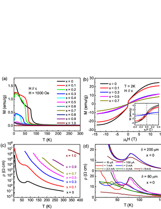
Figure 1 shows the doping effects on magnetization and resistivity of (Mn1-xMgx)3Si2Te6. The temperature dependence of magnetization in Fig. 1(a) reveals a transition at 78 K for Mn3Si2Te6 that is consistent with the reported FIM orderMay et al. (2017); Liu et al. (2018); Martinez et al. (2020); Liu et al. (2021). The FIM order is progressively suppressed with increasing doping until the disappearance for . In addition to the FIM transition, an abnormal decline on the magnetization curve can be observed at low temperatures for , which could be ascribed to a spin reorientation transition due to a doping-induced magnetic dilution (Supplementary Fig. S2)Liu et al. (2023). The saturation magnetization decreases with increasing [Fig. 1(b)]. The hysteresis loops resulting from the FIM can be observed in the low field range in both the undoped and doped samples [see inset in Fig. 1(b)].
Temperature dependence of the resistivity for different compositions is displayed in Fig. 1(c). Anomalies on the resistivity of = 0 and 0.1 samples correspond to the s. When the exceeds 0.1, the anomaly associated with cannot be observed due to the limit of the measured resistance range. The values of the resistivity increase from 10-1 to 107 cm at 300 K in the doping range of . The resistivity for Mg3Si2Te6 is too large to be measured below 328 K in our experimental setupHuang et al. (2023). We fit the resistivity above using the polaron hopping modelLiu et al. (2021) () = exp(a/B), where is the activation energy and is the Bolzman constant. The results reveal linearly increased from 83.5 to 408.6 meV with the Mg2+ doping [Fig. 5], revealing that the gap changes uniformly as doping, which may facilitate the electronic structure tuning in real applications. Figure 1(d) shows the resistivity measurements on Mn3Si2Te6 single crystals with different currents and thicknesses. The can be tuned by the magnitude of current for the single crystal with a thickness of = 80 m, consistent with the previously proposed COC stateZhang et al. (2022a). However, the anomalies in resistivity for the = 200 m sample are hardly changed until the current is up to 8 mA. According to the discrepancy of the s for different currents and samples, the COC state does not depend on the current magnitude but the current density. The critical current density for the FIM state at 0 K is estimated to be 95 mA/mm2 [Supplementary Fig. S1(d)].
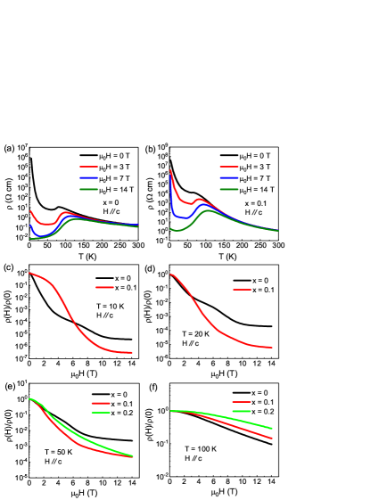
To reveal the doping effect on the CMR, we show the temperature dependence of resistivity with magnetic fields parallel to the axis for the undoped and doped compounds in Figs. 2(a) and 2(b). They all exhibit large CMR below the . Similar measurements on the 0.2, 0.3, 0.5, and 0.7 compositions are shown in the Supplementary Fig. S3. At 5 K, the resistivity of Mn3Si2Te6 drastically decreases by about 8 orders of magnitude from 106 cm at zero filed to 10-2 cm at = 14 T. The change is smaller for (Mn0.9Mg0.1)3Si2Te6 at 5 K. However, over a wider temperature range from 10 K to the , the drop of resistivity for the doped compound under high magnetic fields is larger. To compare the CMR as doping quantitatively, we plot as a function of the magnetic field at 10, 20, and 50 K below the , and 100 K above the in Figs. 2(c-f). Due to the limitation of the measured resistivity range, the data of the compound at 10 and 20 K are not shown in Figs. 2(c) and 2(d). The change of the magnitude of for the doped sample is about 2 orders larger than that of the undoped sample below at 14 T. The magnitudes of reverse below 6.0 T at 10 K, 2.8 T at 20 K, and 1.8 T at 50 K, respectively. The CMR effect becomes much weaker above as shown in Fig. 2(f). Thermal fluctuations, SOC, and thermal activation gap are three ingredients that govern the CMR in (Mn1-xMgx)3Si2Te6. Increasing the temperature below and strengthening the SOC will promote the conductance of electrons as expected. Our results demonstrate that substituting Mn2+ by nonmagnetic Mg2+ will enlarge the thermal activation gap, enhancing the CMR in this system.
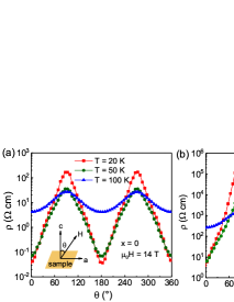
Figure 3 shows a comparison of the colossal AMR for the undoped () and doped () compounds at 20 and 50 K below , and 100 K above . The AMR exhibits a two-fold rotational symmetry. The resistivity as a function of the angle of the magnetic field to the axis evolves close to linearly, consistent with the scenario that the spins of Mn2+ couple with the moments of the COC along the edges of the MnTe6 octahedraZhang et al. (2022a). Upon 10 Mg doping, the thermal activation gap is enlarged, and the magnitude of the AMR in (Mn0.9Mg0.1)3Si2Te6 is enhanced an order at the same measurement conditions. Nonetheless, it is expected that the AMR will be enhanced in a wider doping range and at lower temperaturesSeo et al. (2021).
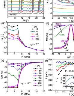
Pressure can effectively tune the structure, electronic transport, and magnetism of layered structural materialsSun et al. (2023a, 2021b); Olmos et al. (2023). Figure 4(a) displays the high-pressure XRD patterns of Mn3Si2Te6 up to 24.2 GPa. A clear structural transition is identified between 13.2 and 14.6 GPa, consistent with the reported Raman spectroscopy measurementsWang et al. (2022). The XRD patterns above 14.6 GPa are consistent with the space group with a monoclinic structure where the lattice constants , Å and at 14.6 GPa. The lattice parameters as a function of pressure can be deduced from the XRD patterns (Supplementary Fig. S4). However, the accurate atomic positions cannot be determined due to the limited reflection peaks.
Under pressure, the resistance measurements reveal a semiconducting to metallic transition [Fig. 4(b)]. Below the pressure of the structural transition, the can be identified as a hump in resistance, yielding a linearly increased from 78 K at ambient pressure to 214 K at 10 GPa. The anomalous Hall effect of Mn3Si2Te6 under pressure is intimately coupled with the magnetism (Supplementary Fig. S5). The thermal activation gap decreases with increasing pressure until closing at around 7 GPa. The activation gaps fitted by the polaron hopping model and the s of Mn3Si2Te6 under pressure have been presented in Fig. 5. The resistance measured at various pressures and temperatures also shows an abrupt drop at 6 GPa [Fig. 4(c)]. Figure 4(d) shows typical magnetoresistance ( defined as (H-0)/100%) measurements at 2.6 GPa and different temperatures (for more pressures, see supplementary Fig. S6). The shows an abrupt change across K at 2.6 GPa [Fig. 4 (d)] and a sharp drop at 6 GPa [Fig. 4 (e)], revealing that the SOC and thermal activation gap are the dominating ingredients that govern the as elevated temperature and pressure, respectively.
The resistance of Mn3Si2Te6 at 20.2 and 26.3 GPa are shown in Fig. 4 (f) to investigate the electronic property of the monoclinic structure. Clear drops can be observed below 5 K on resistance. By applying a magnetic field up to 1.5 T, the resistance at 26.3 GPa below 5 K is enhanced obviously and the drop is finally suppressed, suggesting a superconducting transition. We note that it is common for Te-contained compounds showing superconductivity under pressure, such as WTe2Pan et al. (2015); Kang et al. (2015), CrSiTe3Cai et al. (2020), and EuTe2Yang et al. (2022); Sun et al. (2023b), where the electrons of Te play a dominating role in superconductivity.
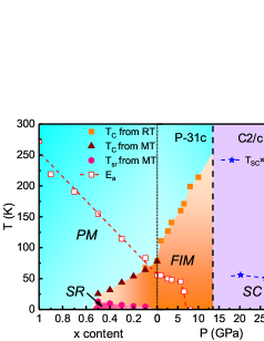
A comprehensive phase diagram combining the electronic, magnetic, and structural measurements as functions of doping and pressure is shown in Fig. 5. The of the FIM transition decreases as doping but increases as applying pressure. The evolution of the thermal activation gap, , reverses, changing from 408.6 meV in Mg3Si2Te6 to 83.5 meV in Mn3Si2Te6 at ambient pressure, and closing at 7 GPa. The magnetoresistance decreases dramatically as the closes, revealing that the CMR of Mn3Si2Te6 is directly coupled with . The presence of magnetoresistance requires SOC that is comparable with the electronic gap. In (Mn1-xMgx)3Si2Te6, the spins of Mn2+ possibly couple with the moments of the COC of the MnTe6 octahedra. The superconductivity observed in the monoclinic structure under pressure may span a large pressure range.
IV SUMMARY
In conclusion, we systematically studied the CMR, AMR, FIM, and structures of (Mn1-xMgx)3Si2Te6 as functions of doping and pressure. As Mn2+ is replaced by Mg2+ progressively, the FIM order is suppressed gradually until disappearing for . For the compositions with between 0.1 and 0.5, a spin reorientation transition appears evidenced by a magnetic anomaly on magnetization at low temperatures. The thermal activation gap is enlarged linearly by the Mg doping. The CMR and AMR are both enhanced in the low-doped samples compared to the parent, which can be attributed to a doping-induced broadening of the band gap and a larger variation range of resistivity. On the contrary, is enhanced with increasing pressure due to the enhanced magnetic exchange interactions until disappearing when undergoing a structural transition above 13.2 GPa. The gap closes and a MIT occurs at 7 GPa, where the CMR is abruptly reduced. A superconducting transition at 5 K in the monoclinic phase under pressure is also observed. Our finding reveals that both doping and pressure are effective methods to change the band structure and magnetism, consequently enhancing the CMR and AMR in the FIM (Mn1-xMgx)3Si2Te6.
V ACKNOWLEDGMENTS
Work at Sun Yat-Sen University was supported by the National Key Research and Development Program of China (grant Nos. 2023YFA1406500, 2023YFA1406002), the National Natural Science Foundation of China (grant No. 12174454), the Guangdong Basic and Applied Basic Research Foundation (grant No. 2021B1515120015), the Guangzhou Basic and Applied Basic Research Funds (grant Nos. 2024A04J6417), and the Guangdong Provincial Key Laboratory of Magnetoelectric Physics and Devices (grant No. 2022B1212010008). We also thank the BL15U1 station in Shanghai Synchrotron Radiation Facility (SSRF) for the help in characterizations.
References
- Grünberg et al. (1986) P. Grünberg, R. Schreiber, Y. Pang, M. Brodsky, and H. Sowers, Physical Review Letters 57, 2442 (1986).
- Baibich et al. (1988) M. N. Baibich, J. M. Broto, A. Fert, F. N. Van Dau, F. Petroff, P. Etienne, G. Creuzet, A. Friederich, and J. Chazelas, Physical Review Letters 61, 2472 (1988).
- Xiao et al. (1992) J. Q. Xiao, J. S. Jiang, and C. Chien, Physical Review Letters 68, 3749 (1992).
- Thompson (2008) S. M. Thompson, Journal of Physics D: Applied Physics 41, 093001 (2008).
- Ennen et al. (2016) I. Ennen, D. Kappe, T. Rempel, C. Glenske, and A. Hütten, Sensors 16, 904 (2016).
- Diény (1994) B. Diény, Journal of Magnetism and Magnetic Materials 136, 335 (1994).
- von Helmolt et al. (1993) R. von Helmolt, J. Wecker, B. Holzapfel, L. Schultz, and K. Samwer, Physical Review Letters 71, 2331 (1993).
- Jin et al. (1994) S. Jin, T. H. Tiefel, M. McCormack, R. Fastnacht, R. Ramesh, and L. Chen, Science 264, 413 (1994).
- Millis et al. (1996) A. Millis, B. I. Shraiman, and R. Mueller, Physical Review Letters 77, 175 (1996).
- Wang et al. (2021) Z.-C. Wang, J. D. Rogers, X. Yao, R. Nichols, K. Atay, B. Xu, J. Franklin, I. Sochnikov, P. J. Ryan, D. Haskel, et al., Advanced Materials 33, 2005755 (2021).
- Shimakawa et al. (1996) Y. Shimakawa, Y. Kubo, and T. Manako, Nature 379, 53 (1996).
- Majumdar and Littlewood (1998) P. Majumdar and P. Littlewood, Physical Review Letters 81, 1314 (1998).
- Rosa et al. (2020) P. Rosa, Y. Xu, M. Rahn, J. Souza, S. Kushwaha, L. Veiga, A. Bombardi, S. Thomas, M. Janoschek, E. Bauer, et al., npj Quantum Materials 5, 52 (2020).
- Yin et al. (2020) J. Yin, C. Wu, L. Li, J. Yu, H. Sun, B. Shen, B. A. Frandsen, D.-X. Yao, and M. Wang, Physical Review Materials 4, 013405 (2020).
- Yang et al. (2021) H. Yang, Q. Liu, Z. Liao, L. Si, P. Jiang, X. Liu, Y. Guo, J. Yin, M. Wang, Z. Sheng, et al., Physical Review B 104, 214419 (2021).
- Sun et al. (2021a) Z. Sun, A. Wang, H. Mu, H. Wang, Z. Wang, T. Wu, Z. Wang, X. Zhou, and X. Chen, npj Quantum Materials 6, 94 (2021a).
- Ogasawara et al. (2021) T. Ogasawara, K.-K. Huynh, T. Tahara, T. Kida, M. Hagiwara, D. Arčon, M. Kimata, S. Y. Matsushita, K. Nagata, and K. Tanigaki, Physical Review B 103, 125108 (2021).
- Vincent et al. (1986) H. Vincent, D. Leroux, D. Bijaoui, R. Rimet, and C. Schlenker, Journal of Solid State Chemistry 63, 349 (1986).
- Ni et al. (2021) Y. Ni, H. Zhao, Y. Zhang, B. Hu, I. Kimchi, and G. Cao, Physical Review B 103, L161105 (2021).
- Seo et al. (2021) J. Seo, C. De, H. Ha, J. E. Lee, S. Park, J. Park, Y. Skourski, E. S. Choi, B. Kim, G. Y. Cho, et al., Nature 599, 576 (2021).
- Sala et al. (2022) G. Sala, J. Lin, A. Samarakoon, D. Parker, A. May, and M. Stone, Physical Review B 105, 214405 (2022).
- Kwon et al. (2023) C. I. Kwon, K. Kim, S. Y. Kim, R. A. Susilo, B. Kang, K. Kim, D. Y. Kim, J. Kim, B. Kim, and J. S. Kim, Current Applied Physics 53, 51 (2023).
- Mijin et al. (2023) S. D. Mijin, A. Šolajić, J. Pešić, Y. Liu, C. Petrovic, M. Bockstedte, A. Bonanni, Z. Popović, and N. Lazarević, Physical Review B 107, 054309 (2023).
- Lovesey (2023) S. W. Lovesey, Physical Review B 107, 224410 (2023).
- Zhang et al. (2022a) Y. Zhang, Y. Ni, H. Zhao, S. Hakani, F. Ye, L. DeLong, I. Kimchi, and G. Cao, Nature 611, 467 (2022a).
- Ye et al. (2022) F. Ye, M. Matsuda, Z. Morgan, T. Sherline, Y. Ni, H. Zhao, and G. Cao, Physical Review B 106, L180402 (2022).
- Zhang et al. (2022b) Q. Zhang, J. Liu, H. Cao, A. Phelan, D. Graf, J. DiTusa, D. A. Tennant, and Z. Mao, NPG Asia Materials 14, 1884 (2022b).
- Zhang et al. (2023) Y. Zhang, L.-F. Lin, A. Moreo, and E. Dagotto, Physical Review B 107, 054430 (2023).
- Sun et al. (2023a) H. Sun, L. Qiu, Y. Han, E. Yi, J. Li, M. Huo, C. Huang, H. Liu, M. Li, W. Wang, et al., Materials Today Physics 36, 101188 (2023a).
- Sun et al. (2023b) H. Sun, L. Qiu, Y. Han, Y. Zhang, W. Wang, C. Huang, N. Liu, M. Huo, L. Li, H. Liu, et al., Communications Physics 6, 40 (2023b).
- Sun et al. (2021b) H. Sun, C. Chen, Y. Hou, W. Wang, Y. Gong, M. Huo, L. Li, J. Yu, W. Cai, N. Liu, et al., Science China Physics, Mechanics & Astronomy 64, 118211 (2021b).
- Cai et al. (2020) W. Cai, H. Sun, W. Xia, C. Wu, Y. Liu, H. Liu, Y. Gong, D.-X. Yao, Y. Guo, and M. Wang, Physical Review B 102, 144525 (2020).
- Prescher and Prakapenka (2015) C. Prescher and V. B. Prakapenka, High Pressure Research 35, 233 (2015).
- Pawley (1981) G. S. Pawley, Journal of Applied Crystallography 14, 357 (1981), ISSN 0021-8898.
- Xu et al. (2018) Y. Xu, W. Li, C. Wang, Z. Chen, Y. Wu, X. Zhang, J. Li, S. Lin, Y. Chen, and Y. Pei, Journal of Materiomics 4, 215 (2018).
- May et al. (2017) A. F. May, Y. Liu, S. Calder, D. S. Parker, T. Pandey, E. Cakmak, H. Cao, J. Yan, and M. A. McGuire, Physical Review B 95, 174440 (2017).
- Liu et al. (2018) Y. Liu, C. Petrovic, et al., Physical Review B 98, 064423 (2018).
- Martinez et al. (2020) L. Martinez, H. Iturriaga, R. Olmos, L. Shao, Y. Liu, T. T. Mai, C. Petrovic, A. R. Hight Walker, and S. Singamaneni, Applied Physics Letters 116, 172404 (2020).
- Liu et al. (2021) Y. Liu, Z. Hu, M. Abeykoon, E. Stavitski, K. Attenkofer, E. D. Bauer, C. Petrovic, et al., Physical Review B 103, 245122 (2021).
- Liu et al. (2023) H. Liu, M. Huo, C. Huang, X. Huang, H. Sun, L. Chen, J. Xu, W. Yin, R. Li, and M. Wang, arXiv:2303.00597 (2023).
- Huang et al. (2023) C. Huang, B. Cheng, Y. Zhang, L. Jiang, L. Li, M. Huo, H. Liu, X. Huang, F. Liang, L. Chen, et al., Chinese Physics B 32, 037802 (2023).
- Olmos et al. (2023) R. Olmos, P.-H. Chang, P. Mishra, R. R. Zope, T. Baruah, Y. Liu, C. Petrovic, and S. R. Singamaneni, The Journal of Physical Chemistry C 127, 10324 (2023).
- Wang et al. (2022) J. Wang, S. Wang, X. He, Y. Zhou, C. An, M. Zhang, Y. Zhou, Y. Han, X. Chen, J. Zhou, et al., Physical Review B 106, 045106 (2022).
- Pan et al. (2015) X.-C. Pan, X. Chen, H. Liu, Y. Feng, Z. Wei, Y. Zhou, Z. Chi, L. Pi, F. Yen, F. Song, et al., Nature communications 6, 7805 (2015).
- Kang et al. (2015) D. Kang, Y. Zhou, W. Yi, C. Yang, J. Guo, Y. Shi, S. Zhang, Z. Wang, C. Zhang, S. Jiang, et al., Nature Communications 6, 7804 (2015).
- Yang et al. (2022) P. T. Yang, Z. Y. Liu, K. Y. Chen, X. L. Liu, X. Zhang, Z. H. Yu, H. Zhang, J. P. Sun, Y. Uwatoko, X. L. Dong, et al., Nature Communications 13, 2975 (2022).